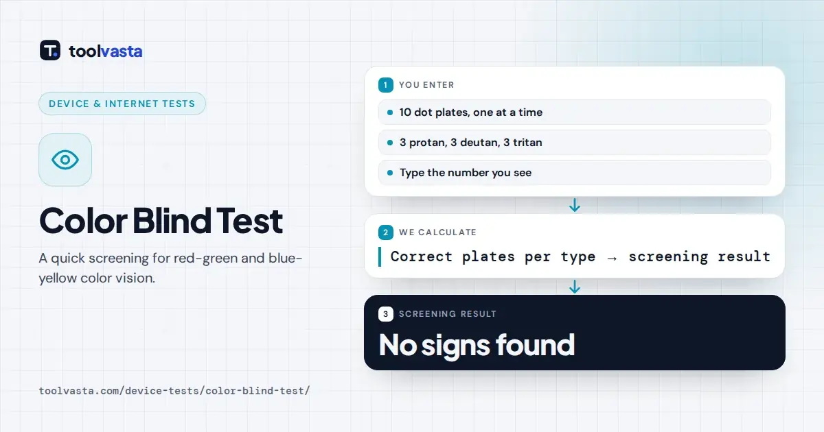
How to Take the Color Blind Test
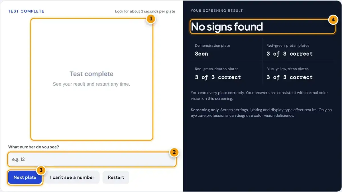
- Look at the dot plate for about 3 seconds on a bright screen.
- Type the number you see in the plate.
- Press Next plate, or I can't see a number if none appears.
- After 10 plates, read your screening result with protan, deutan and tritan scores.
Each of the ten plates is a circle of colored dots, and some dots of a slightly different color form a number. Type the number you see and press Next plate, or simply press Enter.
If nothing stands out, press I can't see a number. Spend about three seconds on each plate rather than hunting for a hidden shape. The first one is a demonstration plate that nearly everyone reads.
Before starting, set full screen brightness and turn off night mode, any blue light filter and color adjustment apps. Then sit about an arm's length away, in normal room light, facing the screen straight on.
What a Color Blind Test Checks
A color blind test, also written colour blind test or colour blindness test, screens for color vision deficiency. It checks whether you can easily tell apart two colors that most people see as clearly different.
Normal color vision relies on three kinds of cone cells in the retina: long, medium and short wavelength cones. When one kind is missing or works weakly, certain pairs of colors start to look alike.
Most people search for a red green color blindness test, since that is by far the most common kind. This test covers red-green deficiency and also includes three plates for the much rarer blue-yellow deficiency.
How These Plates Are Made
The plates are generated by our own code, not copied from any published test. They follow the same pseudoisochromatic idea: the number and the background look alike except for one small, carefully hidden color difference.
For each screening plate, the background color is converted to a cone response using a matrix published by Viénot, Brettel and Mollon in 1999. One cone value is raised, then converted back to screen colors.
number color: L (protan plate), M (deutan plate) or S (tritan plate) raised by k
number RGB = MRGB→LMS−1 × changed cones
Random lightness changes in every dot hide the number from brightness cues. Each type has a strong, medium and faint plate, from 18% to 6.5% for L and M, and 80% to 34% for S.
How Your Result Is Scored
There are three plates for each type: protan for long wavelength cones, deutan for medium and tritan for short. The summary compares the misses in each group to see which pattern your answers follow best.
In a worked example, suppose you read the demonstration plate, every deutan plate and every tritan plate, but miss all three protan plates. The summary would then report a pattern consistent with a protan type.
If you miss only the single faintest plate, the result says mostly normal, because one miss often comes from screen settings. Missing the demonstration plate asks you to check your setup before trusting anything else.
Types of Color Vision Deficiency
Each type of deficiency is named after the cone it affects most. The ending anomaly means the cone works weakly, and the ending anopia means the cone is missing, so protanopia is stronger than protanomaly.
| Type | Cone affected | Colors often confused |
|---|---|---|
| Deuteranomaly / deuteranopia | M (green) | Red, green, brown, orange |
| Protanomaly / protanopia | L (red) | Red, green, and reds look darker |
| Tritanomaly / tritanopia | S (blue) | Blue and green, yellow and violet |
| Achromatopsia | All cones | Sees little or no color |
Red-green deficiency is X-linked, so it is far more common in men. The National Eye Institute reports it affects up to about 1 in 12 men and 1 in 200 women of Northern European ancestry.
Deuteranomaly is the single most common form overall. Tritanopia and tritanomaly are rare and are usually not linked to the X chromosome, and achromatopsia, seeing little or no color at all, is very rare indeed.
Tests Eye Doctors Use
Eye care professionals use several different tests, often together, to confirm the type and severity of a deficiency. Printed plates are viewed under controlled lighting, which removes the screen differences that affect any online version.
- Ishihara plates: the best known plate test, with 38 plates in the full edition, designed for red-green screening.
- HRR plates: grade red-green and blue-yellow deficiency from mild to severe.
- Farnsworth-Munsell 100 Hue and D-15: arrange colored caps in order of hue.
- Anomaloscope: match two halves of a light field, the most precise red-green test.
- Lantern test: identify colored lights, used for some transport and marine jobs.
Ordering tests such as the Farnsworth-Munsell 100 Hue show how finely you separate similar colors. The shorter D-15 version sorts only fifteen caps and is often chosen when a clinic needs a much quicker assessment.
An anomaloscope asks you to mix red and green light until it matches a yellow field. It is the reference test for red-green types, but few clinics have one, so plate tests are used first.
Limits of an Online Test
This test cannot give a diagnosis or measure severity. Phones and monitors show colors differently, and none of them are calibrated like printed clinical plates, so the same eyes can score differently on two screens.
A blue light filter or warm night mode can make the tritan plates hard for anyone. If you miss those plates, turn off every filter and repeat the test before drawing any conclusion from it.
Jobs such as commercial pilots, licensed electricians and some police or military roles require approved, in-person color tests. Only an optometrist or ophthalmologist can provide the formal assessment that employers, schools and licensing bodies accept.
What to Do Next
If the summary suggests a deficiency, book a color vision assessment with an optometrist. A clear diagnosis helps at school, at work, on the road and when choosing a career where color matters for safety.
There is no cure for inherited color blindness, as the MedlinePlus genetics guide explains, but color-correcting apps, labels and high-contrast settings make daily tasks easier. Some tinted lenses also help certain people distinguish some colors.
Color vision can also be acquired later through eye disease, injury or some medications. A sudden change, or a change in one eye only, should be checked promptly because it can signal another eye condition.
Frequently asked questions
Is this online color blind test accurate?
It is a screening test that can point to a likely red-green or blue-yellow deficiency. Screen settings affect results, so it cannot replace an in-person test by an eye care professional.
What does it mean if I cannot see the numbers?
Missing several plates of the same type suggests a color vision deficiency of that type. Check your screen brightness and filters first, then see an optometrist for a proper assessment.
What is the most common type of color blindness?
Deutan types, which reduce sensitivity to green light, are the most common. Together with protan types they make up red-green color vision deficiency, the kind most people mean by color blind.
Can women be color blind?
Yes, but it is much less common. Red-green deficiency is inherited on the X chromosome, so women usually need two affected copies of the gene, while men need only one.
Are these the same plates used by doctors?
No. The plates here are generated by our own software using cone response models. Clinical tests such as Ishihara use printed, calibrated plates viewed under controlled lighting.
Can color blindness be cured?
Inherited color blindness has no cure, and it does not usually get worse. Apps, labels and high-contrast settings help with daily tasks, and an eye exam can check any sudden change.